ANIMAL TISSUES
In multicellular animals, various specialized cells work together to perform vital functions, exhibiting a division of labor that ensures the survival of the entire organism. This organizational hierarchy consists of cells, tissues, organs, and organ systems.
1. Cells:
- Unicellular organisms perform all essential functions, such as digestion, respiration, and reproduction, within a single cell.
- In complex organisms, like humans, these functions are distributed among different cell types, each with a unique role.
2. Tissues:
- Tissue is an organization of similar cells, along with intercellular substances, that work collectively to perform specific functions.
- Complex animals primarily consist of four basic types of tissues: epithelial, connective, muscular, and nervous.
- Organs are formed when various tissues are organized in specific proportions and patterns to serve a specialized purpose.
- Examples of organs in the human body include the stomach, lungs, heart, and kidneys.
- Multiple organs can collaborate to carry out a common function through physical and/or chemical interactions.
- These groups of organs collectively form organ systems, such as the digestive system and respiratory system.
- Within multicellular organisms, the division of labor is essential for efficient functioning and overall survival.
- Cells, tissues, organs, and organ systems work in harmony to carry out distinct tasks, contributing to the well-being of the entire organism.
(i) Epithelial,
(ii) Connective,
(iii) Muscular and
(iv) Neural
- Epithelial tissue, often referred to as epithelium, is a critical component of animal tissues.
- It covers body surfaces and linings, providing protection and facilitating various functions.
- Epithelial tissues can be broadly categorized into simple and compound epithelium, with further differentiation based on structural modifications of the cells.
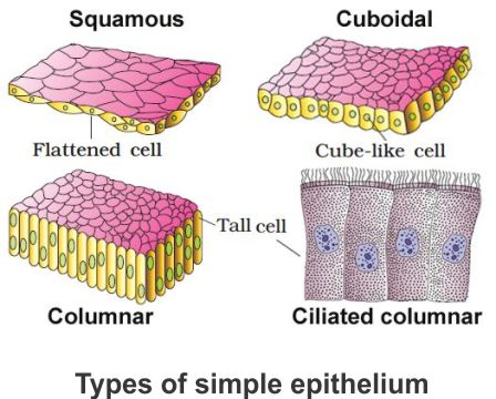
1. Simple Epithelium:
- Simple epithelium consists of a single layer of cells.
- Functions: It serves as a lining for body cavities, ducts, and tubes.
a. Squamous Epithelium:
- Composed of thin, flattened cells with irregular boundaries.
- Found in the walls of blood vessels and air sacs of the lungs, facilitating diffusion.
b. Cuboidal Epithelium:
- Composed of cube-like cells.
- Commonly found in gland ducts and parts of nephrons in the kidneys, involved in secretion and absorption.
c. Columnar Epithelium:
- Composed of tall, slender cells with nuclei located at the base.
- Found in the lining of the stomach and intestines, aiding in secretion and absorption.
d. Ciliated Epithelium:
- Contains cilia on the free surface.
- Function is to move particles or mucus in a specific direction.
- Found in organs like bronchioles and fallopian tubes.
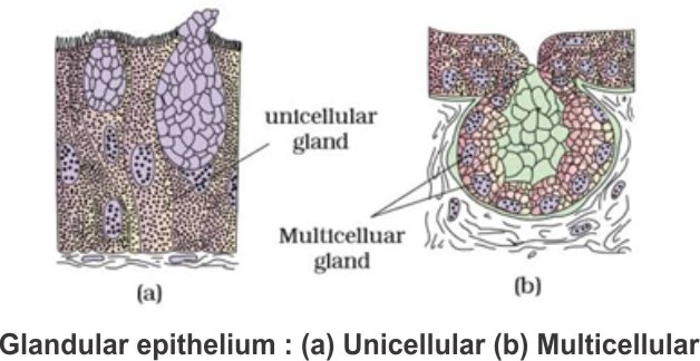
e. Glandular Epithelium:
- Glandular epithelium is a specialized type of columnar or cuboidal epithelial tissue responsible for secretion.
- These glands can be categorized into two main types: unicellular and multicellular.
- Additionally, glands are further divided based on the mode of secretion into exocrine and endocrine glands.
- Glandular epithelium refers to cells specialized for secretion and can be found in columnar or cuboidal forms.
- Glandular epithelium can be classified into two main types based on the structure and organization of the gland cells.
a. Unicellular Glandular Epithelium:
- Unicellular glands consist of isolated glandular cells.
- Example: Goblet cells found in the alimentary canal, which secrete mucus to protect and lubricate the digestive tract.
- Multicellular glands are formed by clusters of glandular cells working together.
- Example: Salivary glands, which secrete saliva to aid in digestion.
Classification Based on Secretion Mode:
Glands can be further categorized based on how they release their secretions.
a. Exocrine Glands:
- Exocrine glands secrete their products into ducts or tubes that lead to specific target locations.
- Common exocrine gland products include mucus, saliva, earwax, oil, milk, and digestive enzymes.
- The secretions are released at the body's surfaces or into cavities.
b. Endocrine Glands:
- Endocrine glands differ from exocrine glands in that they do not have ducts.
- Instead, they secrete hormones directly into the fluid bathing the gland.
- These hormones are then transported through the bloodstream to distant target organs and tissues, regulating various physiological processes.
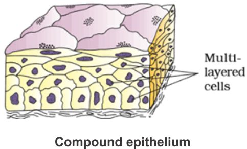
2. Compound Epithelium:
- Consists of more than one layer of cells.
- It has a limited role in secretion and absorption.
- Mainly provides protection against chemical and mechanical stresses.
- Locations include the skin's dry surface, the moist surfaces of the buccal cavity, pharynx, inner linings of salivary gland ducts, and pancreatic ducts.
Cell junction in Epithelial Tissue
- Cell junctions are specialized structures that play a crucial role in maintaining the integrity and functionality of epithelial tissue, as well as other animal tissues.
- These junctions provide both structural support and enable cellular communication.
- There are three main types of cell junctions: tight junctions, adhering junctions, and gap junctions.
- Function: Tight junctions are essential for creating a barrier that prevents the leakage of substances across the tissue.
- Structure: These junctions consist of proteins that form a continuous, impermeable seal between adjacent cells.
- Role: Tight junctions maintain the selective permeability of the epithelium, controlling the passage of ions, molecules, and preventing the uncontrolled movement of substances.
- Function: Adhering junctions perform a "cementing" function, keeping neighboring cells together.
- Structure: These junctions are formed by protein complexes that link cells and anchor them to the underlying cytoskeleton.
- Role: Adhering junctions provide mechanical stability to tissues and allow for coordinated movement, especially in tissues that experience stretching or bending.
3. Gap Junctions:
- Function: Gap junctions enable direct communication between neighboring cells by connecting their cytoplasm.
- Structure: Gap junctions are composed of connexin proteins that form channels called connexons, allowing the passage of ions, small molecules, and even larger molecules.
- Role: These junctions facilitate rapid transfer of signals, nutrients, and other essential substances between cells, ensuring coordinated functions in the tissue.
- Connective tissues play a vital role in the complex animal body, where they are highly abundant and widely distributed.
- Named for their primary function of connecting and supporting various organs and tissues, connective tissues range from softer varieties to specialized types, including cartilage, bone, adipose tissue, and blood.
- In nearly all connective tissues (except blood), cells secrete fibers composed of structural proteins, such as collagen or elastin.
- These fibers are crucial for providing strength, elasticity, and flexibility to the tissue. Additionally, connective tissue cells secrete modified polysaccharides, which accumulate between cells and fibers, forming a matrix or ground substance.
- Connective tissues can be classified into three primary types:
i) Loose connective tissue,
ii) Dense connective tissue and
iii) Specialised connective tissue
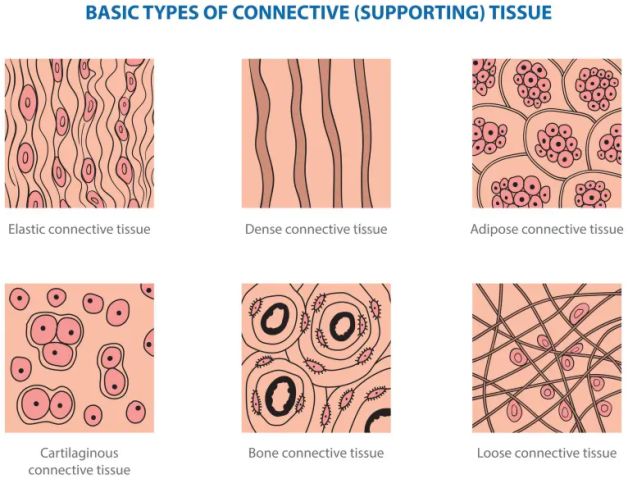
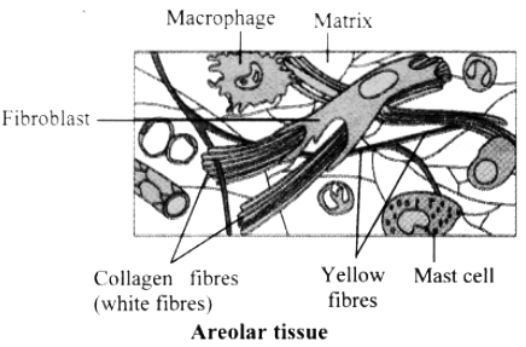
1. Loose Connective Tissue
- Loose connective tissue is a versatile type of connective tissue characterized by the loose arrangement of cells and fibers within a semi-fluid ground substance.
- This tissue provides support for epithelium and plays a crucial role in maintaining the structural integrity of various body structures.
- Structure: Loose connective tissue features a semi-fluid matrix in which cells and fibers are loosely arranged.
- Function: It acts as a supporting framework for epithelial tissues, offering flexibility and support.
- Components:
a. Fibroblasts: Cells responsible for producing and secreting fibers.
b. Macrophages: Immune cells that help defend the body against pathogens.
c. Mast Cells: Cells involved in the immune response, particularly allergic reactions and inflammation.
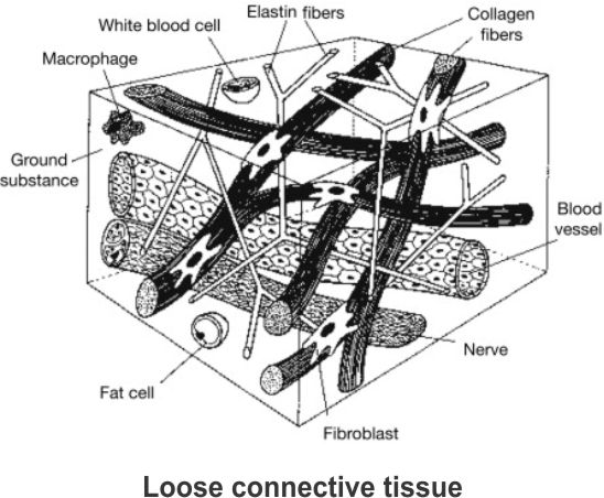
- One specific example of loose connective tissue is the areolar tissue. Areolar tissue is a common type of loose connective tissue found beneath the skin, connecting and supporting various structures in the body.
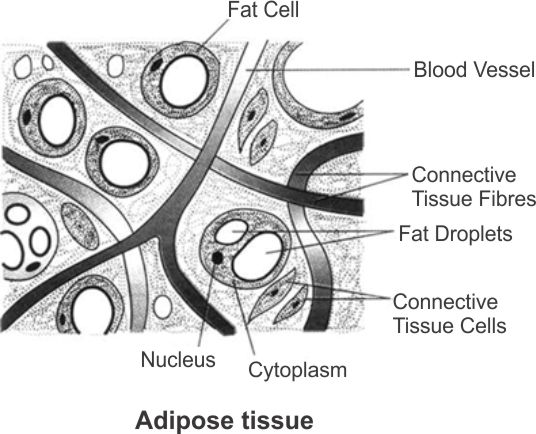
- Additionally, adipose tissue is another type of loose connective tissue primarily located beneath the skin, with cells specially adapted for fat storage.
- Structure: Adipose tissue consists of specialized cells known as adipocytes that are designed for fat storage.
- Function: Its primary function is to store excess nutrients in the form of fats. This stored energy can be used when needed.
- Specialization: Adipocytes have a unique structure adapted for fat storage, with a single large lipid droplet occupying most of the cell's volume.
- Location: Adipose tissue is mainly found beneath the skin (subcutaneous) and around internal organs (visceral), providing insulation and cushioning, as well as serving as an energy reservoir.
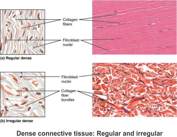
2. Dense Connective Tissue
- Dense connective tissues are a specialized category of connective tissues characterized by their compact arrangement of fibers and fibroblasts.
- These tissues are classified into two main types based on the orientation of fibers: dense regular and dense irregular.
- Each type serves specific functions within the body and is found in distinct locations.
- Structure: In dense regular connective tissues, collagen fibers are neatly arranged in rows, forming a regular pattern.
- Function: These tissues provide strong and durable connections between structures that require unidirectional tensile strength.
- Examples:
a. Tendons: Attach skeletal muscles to bones, allowing the transmission of muscular forces to enable movement.
b. Ligaments: Connect one bone to another, providing stability and support to joints.
- Structure: Dense irregular connective tissue is characterized by its fibroblasts and the presence of many fibers (primarily collagen) that are oriented in various directions.
- Function: This tissue offers strength and support while accommodating multidirectional stresses.
- Location: Dense irregular connective tissue is primarily found in the skin and other areas where tissue integrity and flexibility are required.
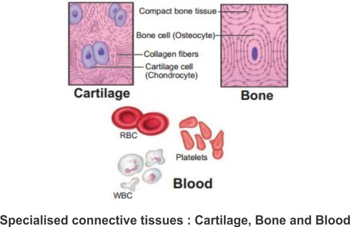
3. Specialised Connective Tissue
- Specialized connective tissues play critical roles in the complex animal body, serving unique functions that contribute to structural support, resilience, and the transportation of vital substances.
- This category includes cartilage, bones, and blood, each characterized by distinct features and functions.
a. Cartilage:
- Structure: Cartilage has solid and pliable intercellular material that resists compression.
- Cells: Chondrocytes, the cells within cartilage, are enclosed in small cavities within the matrix secreted by them.
- Functions:
- Cartilage provides structural support and resilience.
- It resists compression, maintains the shape of various body structures, and allows flexibility in joints.
- Examples of Locations: Cartilage is present in the tip of the nose, outer ear, joints, between adjacent bones in the vertebral column, limbs, and hands in adults.
- Structure: Bones have a hard and non-pliable ground substance rich in calcium salts and collagen fibers, contributing to their strength.
- Cells: Osteocytes are the bone cells present in small spaces called lacunae.
- Functions:
- Bones form the primary structural framework of the body.
- They support and protect softer tissues and organs.
- Bones interact with skeletal muscles to facilitate movements.
- Some bones contain bone marrow, where blood cells are produced.
- Examples of Locations: Limb bones, such as the long bones of the legs, serve weight-bearing functions and enable mobility.
- Composition: Blood is a fluid connective tissue consisting of plasma, red blood cells (RBCs), white blood cells (WBCs), and platelets.
- Functions:
- Blood is the primary circulating fluid responsible for transporting essential substances throughout the body.
- Red blood cells transport oxygen, while white blood cells play a crucial role in the immune response.
- Platelets are essential for blood clotting and wound healing.
- Blood plasma carries various solutes and nutrients, contributing to homeostasis and overall health.
- Muscles are essential components of the human body responsible for generating movement and maintaining various bodily functions.
- These contractile tissues are composed of long, cylindrical fibers arranged in parallel arrays.
- Within these muscle fibers, numerous fine fibrils, known as myofibrils, facilitate the process of contraction and relaxation.
- The coordinated actions of muscles play a pivotal role in adapting to environmental changes and maintaining the positions of different body parts.
- Muscles are of three types, skeletal, smooth, and cardiac.
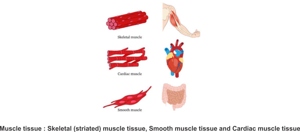
1. Skeletal Muscle
- Skeletal muscle tissue is a critical component of the musculoskeletal system.
- Attachment to Bones: Skeletal muscle tissue is closely attached to skeletal bones, allowing for the transmission of muscular forces to the bones and facilitating controlled movement.
- Organization: In a typical skeletal muscle, such as the biceps, individual skeletal muscle fibers are bundled together in a parallel arrangement.
- Striated Appearance: Skeletal muscle fibers are striated, displaying a striped appearance due to the repeating arrangement of actin and myosin protein filaments.
- Connective Tissue Sheath: A protective sheath of tough connective tissue encloses several bundles of muscle fibers within a muscle.
- This type of muscle is typically found in the limbs and plays a fundamental role in producing voluntary movements.
- Fusiform Shape: Smooth muscle fibers have a tapered, fusiform shape, meaning they are narrower at both ends.
- Lack of Striations: Unlike skeletal muscle, smooth muscle fibers do not display striations, which are the repeating dark and light bands seen in striated muscle.
- Cell Junctions: Cell junctions, including gap junctions, hold smooth muscle fibers together and allow for coordinated contractions.
- Connective Tissue Sheath: Bundles of smooth muscle fibers are typically enclosed in a connective tissue sheath, providing support and organization.
- Location-
- Internal Organs: Smooth muscle is primarily found in the walls of internal organs such as the blood vessels, stomach, intestines, and various other structures.
- Involuntary Function: Smooth muscles are considered "involuntary" because their functioning is not directly controlled by conscious thought. Contractions of smooth muscles are typically regulated by the autonomic nervous system and hormonal signals.
- Exclusive to the Heart: Cardiac muscle tissue is found exclusively in the heart, where its contractions are essential for pumping blood throughout the circulatory system.
- Cell Junctions: Cardiac muscle cells are held together by cell junctions, particularly intercalated discs. These junctions fuse the plasma membranes of adjacent cells.
- Intercalated Discs: Intercalated discs are specialized communication junctions located at specific fusion points between cardiac muscle cells.
- Coordinated Contraction: Intercalated discs facilitate communication between cardiac muscle cells. When one cell receives a signal to contract, the intercalated discs ensure that neighboring cells are also stimulated to contract.
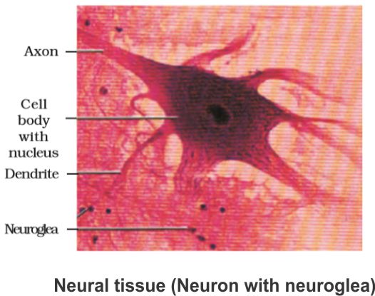
- Neural tissue plays a fundamental role in controlling the body's responses to changing conditions and stimuli.
- At the core of the neural system are neurons, excitable cells that enable the transmission of electrical signals.
- Neurons:
- Excitable Cells: Neurons are the basic functional units of the neural system and are excitable, meaning they can generate and transmit electrical signals.
- Transmission of Electrical Disturbances: When appropriately stimulated, neurons generate electrical disturbances that swiftly travel along their plasma membranes.
- Output Zone: These electrical disturbances, upon reaching the neuron's endings or output zone, trigger events that can stimulate or inhibit adjacent neurons and other cells.
- Supporting the neurons are neuroglial cells, which make up a significant portion of the neural system, providing protection and support to the neurons.
- Neuroglial Cells:
- Supportive Role: Neuroglial cells constitute the rest of the neural system and serve a vital role in protecting and supporting neurons.
- Abundance: In the human body, neuroglia make up more than half of the total volume of neural tissue, emphasizing their significance in neural function.
- These cells collectively govern the body's responsiveness to various stimuli, and the transmission of electrical disturbances within neurons is a key mechanism underlying neural function.